

Buyer's Guide
Long-Length, Full-Spine, and Long-View DR
2026-10-30 · updated 2026-11-05 · long-length DR · full spine · scoliosis
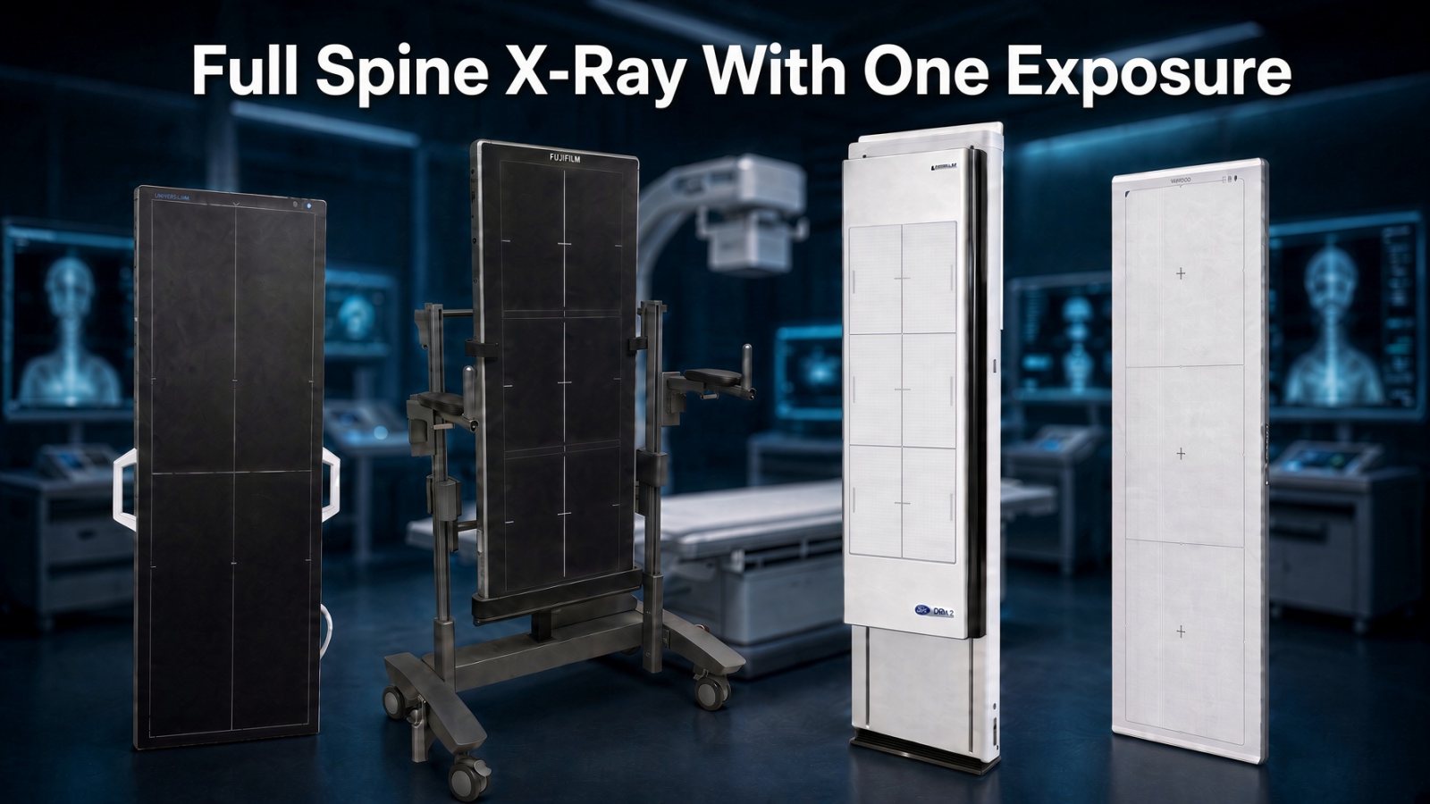
Full spine with one exposure. The labels you can read on this lineup are Universal, Fujifilm, Del Medical, Medicatech, and Delworks LLI. Each system is photographed again in its own section below.
A full spine is not only a chiropractic study. Orthopedic and pediatric spine practices, neurosurgery, physiatry, and osteopathic physicians use a long film for scoliosis and a standing spine, and so do the imaging centers that shoot for them. Chiropractic is where the one-exposure long panel became everyday equipment, and the 2020 Imaging system is built with that measurement work in mind. Six detectors belong in the same conversation. Four of them were already here. Medicatech’s Venu 1748 and the Delworks LLI are the two added from their current brochures.
If you want the short version: for most chiropractic full-spine rooms I start with the 2020 Imaging Universal 17×48. One exposure, a preview in under six seconds, and the measurement tools you already use. Fujifilm, Vieworks, iCRco, Medicatech, and Delworks each have a real place in this conversation. The notes below are there so you can see what each one does well, and tell me what sounds like your room.
Clinics ask for this four ways. A long-length panel. A full-spine detector. A long-view image. A long-form receptor. In an upright room those words describe the same job: the whole spine on one image. Stitching separate 17×17 views is still a great option for most chiropractors, and many offices stay with it for good reason. A long panel is the other path, one exposure, when that is what you want.
Four of these systems were already a fair set, and each one still earns its place. Two more belong beside them. 20/20 Imaging 17×48 is where a typical chiropractic full-spine room starts. Fujifilm FDR D-EVO GL is one of the original long-length lines, and it firmly deserves its spot here. Vieworks 17×51 offers the longest imaging area on this page, 17 by 51 inches, so more of a tall patient fits on a single image. iCRco iDR-L publishes excellent resolution numbers on a scanning system. That is an earlier technology than a flat panel. Medicatech Venu 1748 is a cesium full-spine and long-bone panel, wired or Wi-Fi, with a rolling cart. Delworks LLI is the lighter 17×42 panel, built to leave the wall. A dash means that figure was not published. It is not a zero.
Quotes stay private. Call 720-600-3117. Tell me what you already have, and what you want the room to do.
How long-length imaging got here
Del Medical published a white paper in June 2021, document #9000-LLI-WP, Rev. A, called “Long Length Imaging.” The timeline below is from that paper. Where they make a claim about cost, or about being first, I am leaving it as their claim. I have not measured those myself.
Film
The largest everyday film was 14×17. A full spine or a hip-to-ankle study took more than one cassette. A scanogram was often three separate 14×17 exposures — pelvis, knee, and ankle — with the patient supine and a ruler between the legs. If the patient moved between exposures, the measurement moved with them. Another method overlapped cassettes under one exposure. Those cassettes sat at different distances from the tube, so magnification was not the same on each film. A later film method used one 14×36 cassette. Some of that film was trifold, so it stored in a standard jacket.
Computed radiography
CR started at the same 14×17 size. When departments wanted to leave film, CR copied the old methods. Overlapping cassettes, up to four, covered a field as long as 17×52, and the reader’s software corrected for the magnification change. Or one phosphor plate, up to 14×33, was scanned in parts and stitched. The image could take several minutes.
Early digital radiography
DR made the image on the detector instead of a cassette. Del notes that US payers put about a 7 to 20 percent reimbursement penalty on CR and film exams, which is what pushed a lot of departments off those systems. For years the largest DR plate was still 17×17. Departments either kept a CR reader just for the long studies, or they shot several DR images and stitched them. Cycle time came down from 15–20 seconds toward 1–5 seconds. That is what made a robotic room practical: the tube and the receptor move, then the software stitches.
Most of those rooms rotate the tube around one focal spot, so the overlap looks more like the old film method. A parallel move keeps the tube and the detector facing each other and shifts the focal spot, which Del says makes the overlap harder to stitch. Del also says a fully robotic room built for that work is often as much as three times the cost of a manual room. That is their comparison, not a number from me. Acquiring the set could take 40 seconds or more, and the patient had to hold still the whole time. Density also changes from one exposure to the next, and the blend in the overlap can soften fine detail.
Overlapping several DR plates inside one housing was another path. The plates cannot sit edge to edge, because of the electronics at the border, so software has to hide the seam. One exposure takes patient motion out of the exam. Those panels were heavy, and they were hard to move from the wall to a table or into surgery.
One detector, and the stitching room that stayed
In 2019 Del Medical launched the DELWORKS LLI. Their paper calls it the world’s first monolithic, meaning single-piece, long-length DR detector, at 17×42 inches. I am leaving “world’s first” as their statement. What matters in a room is what the panel does: one exposure, no overlap dose, and a weight you can actually carry. That 2021 paper says the image was ready in 9 seconds and the panel weighed 24 pounds at 0.8 inches thick. The current specification table, brochure #9000-DELWORKSLLI Rev. C, lists 22 pounds and a 3-second acquisition. The matrix uses the current table. The 9-second figure stays as their display time, not as a second acquisition time.
In 2021 Del also introduced the OTC18S. That is not another long panel, and it is not a column here. It is a room that still uses a standard-size detector, shoots more than one exposure, and stitches, with motorized tube rotation. It is for a practice that wants automated stitching without buying a full robotic suite. I mention it so the history is complete.
Two layout points from that paper apply to every long panel on this page, not only Del’s. They recommend a source-to-image distance (SID) of about 100 inches (254 cm) when you want the whole detector covered, and less change in magnification from the center of the spine to the ends. A shorter distance, which is common in surgery, covers less of the panel. And even though DR has more gray scale than film, attenuation filters are still worth using on a scoliosis or a leg-length view. The neck and the ankles do not need the same exposure as the hips. One exposure is not a reason to ignore the thin parts.
Panel matrix
Six columns, in equal width: 2020 Imaging, Vieworks, Fujifilm, iCRco, Medicatech Venu 1748 (the full-spine and long-bone detector), and Delworks LLI. Scroll sideways on a phone. The holder and grid rows for the 2020 come from that quote. Medicatech’s numbers are from the Venu 1748 brochure. Del’s numbers are from brochure #9000-DELWORKSLLI, Rev. C, and where that brochure disagrees with itself I say which line I used. Choose a term and it opens the note below. Each note can return you to that same row, so you do not have to scroll back to the top. A dash means that figure was not published for that system. It is not a zero.
What those words mean for your room
The first bullet is the point. The second is there if you want the reason. Use Back to matrix when you are ready for the next term.
Type
Back to matrix- A flat panel makes the image in one exposure. A scanning system reads the plate after the exposure.
- The 2020, Vieworks, Fujifilm, Medicatech Venu 1748, and Delworks LLI detectors are amorphous-silicon flat panels. The iCRco iDR-L is a scanning phosphor-plate system. Both approaches cover a long spine. The difference is how the image is made and how the room is built around it.
Active Area (inches)
Back to matrix- This is how much of the patient you actually see on the image.
- A longer area is what keeps the skull or the sacrum from being cut off on a tall patient. Vieworks is 51 inches, iCRco’s scan area is 51, Fuji is 49, and the 2020 is 48. Medicatech publishes 425 × 1208 mm, which is about 16.7 × 47.6 inches. Delworks is 17 × 42. Forty-eight inches covers most chiropractic full-spine work, which is why I start there. Forty-two inches is a real step down on a tall standing spine, and it is also why Del’s panel can do a routine view and still leave the wall. Extra length is a real advantage when the patient is tall. Length is one part of the choice, not the only one.
Active Area (mm)
Back to matrix- The same field of view, in millimeters.
- You only need it if you want to check the pixel math yourself. That arithmetic is in the section below.
Pixel Pitch (µm)
Back to matrix- How far apart the pixels are. A smaller number can hold finer detail.
- 139 and 140 microns are where these modern long-length flat panels sit. Fujifilm is published at 150 microns, a little larger. Medicatech is 139. Del’s specification table is 139; the highlights box on the same brochure says 140. I use 139. iCRco’s 100-micron figure is its finest scanning mode, and that mode takes longer to read.
Nyquist / Spatial Resolution (LP/mm)
Back to matrix- The finest detail the detector can theoretically record. Higher is finer.
- 3.5 to 3.6 line pairs is where the 2020, Vieworks, and Delworks are published. Medicatech publishes 3.4 lp/mm. The arithmetic from a 139-micron pitch is 3.6, so their published spatial-resolution number sits a little under the Nyquist figure. I did not change their number to match the math. Fujifilm is 3.3. iCRco publishes 5.0 in its finest scan mode, and that is a strong number. A published line-pair figure and the image on the reading station are worth looking at together, on every system.
Pixel Matrix (full)
Back to matrix- How many pixels are in the finished image.
- More pixels means more data, not automatically a better diagnosis. It is active area divided by pixel pitch. The 2020 matrix is not printed on the sheet. The math lands on 3064 × 8696. Medicatech does not print a matrix either. Del prints 7680 × 3072, which is the long side first. The highlights box lists 3072 × 7680.
Bit Depth
Back to matrix- How many shades of gray the detector can separate. 16-bit is 65,536 shades.
- All of them except Medicatech print 16-bit. That is 65,536 shades, and it is enough gray scale for diagnostic work. Medicatech does not print bit depth on the brochure I used, so that cell is a dash. It is not a zero, and it is not a guess.
Image Preview Time
Back to matrix- How long you and the patient wait before a usable image is on the screen.
- The 2020 is under six seconds. Medicatech’s pre-image is 3 seconds. Del’s specification table says 3 seconds to acquire. The same Del brochure’s highlight, and Del’s 2021 white paper, say about 9 seconds until the image is on screen. I am keeping both numbers and saying which document each one comes from. Fuji’s full-length preview is 9 to 20 seconds. The iCRco is about 55 seconds while the scanner travels the plate. Those are different workflows, and each one is a fit for a different room.
Full Image / Cycle Time
Back to matrix- How long until the full image is ready and you can take the next view.
- Vieworks is about 8 seconds to acquire and 15 before the next shot. Medicatech’s full image is 8 seconds. Fuji’s full-length cycle is about 26 seconds. The iCRco stays near 55. Del’s spec table does not list a cycle beyond the 3-second acquisition. Cycle time is part of how the day flows. It is one reason among several.
Weight
Back to matrix- What the detector, or the whole stand, weighs.
- In a room where the detector stays in the wall stand, weight rarely decides the purchase. Vieworks and Fuji are both about 43 pounds. Medicatech is 17 kg wired, about 37.5 pounds, and 18 kg with the Wi-Fi battery, about 39.7 pounds. Del’s current specification table is 22 pounds (10 kg) with the battery. Their June 2021 white paper said 24 pounds. I use the current sheet in the matrix. The iCRco wall stand is about 100 pounds, because you are looking at the whole stand. The two lighter panels are the ones built to leave the wall.
Dimensions
Back to matrix- Whether the detector physically fits the stand and the wall you already have.
- The wall and the stand are measured with you before anyone says a detector will fit. Medicatech’s outer size is 1271 × 487 × 21 mm. Del’s specification table is 111.5 × 47.1 × 1.5 cm. The drawing on that same page marks the panel at about 44 × 18 × 0.8 inches. I am not averaging those two Del figures.
Scintillator
Back to matrix- The material that turns the X-ray into light the detector can record.
- Cesium iodide (CsI) uses the exposure efficiently, and it is the material I look for on a long panel. The crystals grow like needles, so the light stays in a narrow path to the pixel. GOS and Gadox are powders. Light scatters sideways in the powder, so some of the exposure turns into blur and you generally need more dose for the same clarity. Fujifilm uses GOS and recovers a good part of that with ISS, which reads the light from the side the X-ray hits. Medicatech is CsI. Del’s specification table says Cesium. One paragraph on the features page of that brochure says Gadox, and the next sentence says Cesium Iodide. The matrix follows the specification table, not the mixed paragraph. The quote names which material you are looking at, because it changes dose.
Data Interface
Back to matrix- How the image leaves the detector and gets to the computer.
- A tethered high-speed cable is a natural fit for a stand that stays on the wall. Fujifilm also uses its power supply and a cable run. The iCRco uses a trigger cable so the generator and the scanner work together. Medicatech’s wired model is GigE. The Wi-Fi model is GigE or Wi-Fi. Del is GigE and 802.11ac Wi-Fi, with a 15-foot magnetic cable when you want it wired. That cable also charges the panel.
Trigger Method
Back to matrix- How the detector knows you just exposed.
- Automatic exposure detection works with most generators already in the room, without a new cable. A hard trigger is a cable between the generator and the system, and it is a normal part of some installs. Medicatech lists a software trigger, with AED as an option. Del uses AED and states that the panel works with any generator. Generator compatibility is still confirmed with you so the quote matches the equipment you already have.
Wireless Option
Back to matrix- Whether the detector can work without a data or power cable.
- The 2020, Vieworks, Fujifilm, and iCRco long systems on this page are cabled. A cable is a natural fit when the detector lives in a wall stand. Medicatech offers both: the 1748V is wired, and the 1748V-WF is the Wi-Fi version. Del is wireless, or wired on the charging cable. Wireless is what makes a long panel practical to move from the wall to a table or a cart.
X-ray Range
Back to matrix- The kVp range the detector is specified for.
- Where it is published, 40 to 150 kVp covers normal diagnostic spine work. Vieworks and Delworks both print that range. The others do not, on the sheets I used.
Power
Back to matrix- What has to be plugged in at the stand.
- Fujifilm uses its own power supply, so the outlet at the stand is confirmed before the panel ships. Medicatech lists 27 watts. The Wi-Fi model’s battery is specified at 3 hours on a 90-second cycle. The wired model has no battery. Del lists 20 watts, an adapter from 100–240 volts down to 24 volts at 60 watts, and two lithium-ion batteries: 2 to 3 hours to charge, 650 exposures, 11 hours on standby. The others use a simpler power connection. Either way, the quote says what the room needs.
Operating Environment
Back to matrix- The temperature and humidity the detector is rated for.
- A normal clinical room is inside the ranges that are published. Del lists IPX3, which is a splash rating, and does not print temperature or humidity on this sheet. This is not a reason to pick one panel over another.
Warranty
Back to matrix- How long the manufacturer stands behind the panel.
- The 2020 panel carries a 60-month manufacturer warranty. Warranty for the others was not on this sheet, so it is written on the quote.
Chiropractic Software
Back to matrix- Whether the tools on the screen are the ones you already use on a film or a tracing.
- Symmetry-Sync, which many of you still call Opal-RAD, is the set I trust for this work: Gonstead, Blair, Orthospinology, Cobb, Penning, labeling, and stitching when a study calls for it. Stitching is still a great option for most chiropractic offices. Voyance is Medicatech’s acquisition software. DELWORKS 3.0 is Del’s, and it does include measurement tools. Neither brochure names Gonstead, Blair, or the rest of that chiropractic set. The other panels can be paired with chiropractic software too. That choice is part of the conversation.
Special Technologies
Back to matrix- The manufacturer’s own processing, dose tools, or scan modes.
- On the 2020, REALISM holds trabecular detail without asking the tube for a heavier exposure. Fuji’s ISS, Virtual Grid, and Dynamic Visualization are a real reason an existing Fuji room stays with Fuji, and a real reason to look at Fuji if the room is new to it. Vieworks can take a 17×34 or 17×17 image on the same panel without pulling it out of the stand. Medicatech’s brochure is a single exposure for the full spine or the whole lower extremity, with optional iGrid. Del is also one exposure. You can collimate down for a routine view and DELWORKS auto-shutters to that area. Del’s optional filters sit in the collimator rails for a lateral cervical, an AP or PA scoliosis, or a leg-length view.
Generator Compatibility
Back to matrix- Whether the panel will fire with the generator you already own.
- Most automatic-exposure panels work with generators already in the room. The iCRco is designed around a trigger cable, which is a planned part of that install. Medicatech’s AED is listed as optional, so the quote has to say whether this generator needs it. Del states the LLI works with any generator through AED. I still confirm that against the generator you have.
Manufacturer Location / Support Base
Back to matrix- Who answers the phone on the day the room is down, and where the part ships from.
- 2020 is in Crystal Lake, Illinois. iCRco is in Goleta, California. Fuji’s US office is in Lexington. Vieworks supports the US from Vernon Hills and builds in South Korea. Del Medical is in Harrison, New York, and Bloomingdale, Illinois. Medicatech USA has been supporting products since 2005. The brochure lists a toll-free number and a 949 phone number, and it does not name a city, so I am not going to invent one. The panel in their photo is marked iRay. Medicatech is who the brochure puts in front of the customer. A closer shipping address often means a shorter down day, and every one of these companies can be reached in the US.
Full-Spine Holder
Back to matrix- What holds the detector on the wall, and whether you can move it for the patient in front of you.
- The 2020 quote includes the wall-mounted Trans-Former™ cabinet: 25 inches of vertical travel and a rotating pivot clutch that turns the panel 90 degrees. Holder details for Fuji and Vieworks were not on this sheet, so they are confirmed with the quote. The iCRco is the wall stand itself. Medicatech’s floor stand is the DR-1748D: the detector travels vertically from 1300 to 1770 mm and horizontally from 575 to 1375 mm, the tray turns 90 degrees, and the casters unfold 30 degrees. Del’s fixed upright is the VS50. It hugs the wall, under 7 inches from the wall to the receptor, mounts with no floor drilling, needs no power, and has a lead backer. Vertical travel on the VS50 is 37.5 inches.
Grid
Back to matrix- The grid cleans scatter before it reaches the detector. That is what keeps a long-spine image from looking gray.
- The Trans-Former uses a removable 8:1, 103-line parallel grid. Parallel does not lock you to one source-to-image distance (SID) the way a focused grid does. A focused grid is sharpest at one set distance. Parallel stays useful as that distance changes, which helps when the spine is this long. Del’s optional grid is the same idea: 17×42, 8:1, 103 lines, focal range 44 inches to infinity, so one grid can sit in a routine view or a long-SID spine view. Medicatech lists optional iGrid software and does not print a physical grid ratio on this brochure. Grid details for Fuji, Vieworks, and iCRco were not on this sheet.
Portable / Mobile Stand
Back to matrix- Whether the detector can leave a fixed wall cabinet and still be held for an upright, a table, or a cross-table view.
- Medicatech offers a rolling cart, the DR-1748A, and a floor stand, the DR-1748D. Del offers two mobile holders plus the VS50 wall stand. The Mobile Positioning Holder does upright, supine under-table, and cross-table work, including the operating room. The Mobile Vertical Holder lowers to 1.75 inches from the floor and turns the panel from portrait to landscape. A supine cover on the Del panel is rated at 500 pounds. The 2020 holder on this sheet is the wall Trans-Former, not a rolling stand. The iDR-L is the wall stand. Fuji and Vieworks do not publish a portable long-length stand on the sheets this page uses, so those cells are not a no. They are unpublished here.
Best For
Back to matrix- The short version, after everything above.
- 2020 when the chiropractic tools and a single-exposure flat panel are the priority. Fuji when the room is already Fuji, or when the dose tools are what you want. Vieworks when you want the longest imaging area, and the shorter views on the same panel. Medicatech when you want a cesium long panel with a wired or Wi-Fi choice and a rolling cart. Delworks when the panel has to leave the wall, do a routine view as well as a long view, and stay light enough to carry. iCRco when a US-made scanning wall stand is the way you want to make the image.
ALARA and long-length panels
ALARA means As Low As Reasonably Achievable. It is a radiation protection principle from the International Commission on Radiological Protection, not a certification any panel carries. Three levers matter in practice: time, distance, and shielding. In a full-spine room that becomes exposure time, source-to-image distance, collimation, and attenuation filters on the neck and ankles.
For detectors, ALARA shows up in two measurable specs. Detective quantum efficiency tells you how efficiently the panel converts X-rays into image signal — higher DQE means less exposure for the same image. Scintillator type matters too: cesium iodide generally outperforms gadolinium oxysulfide on dose. Single-exposure geometry is the third lever. Stitching three 17-by-17 images means three exposures, repositioning, and overlap zones that get re-radiated. One tall detector eliminates all three.
Digital also creates a failure mode called dose creep. Unlike film, an overexposed digital image still looks acceptable, so technique quietly drifts up over months. The panel will not stop you. A documented technique chart, periodic exposure-indicator audits, and collimation to anatomy are what keep a room ALARA in practice. Colorado's Part 4, Section 4.5.2 requires facilities to maintain ALARA procedures, and Part 6, Appendix 6F asks for the policy during inspection.
This is educational information, not legal, regulatory, or medical advice. Confirm current Colorado requirements with the CDPHE X-Ray Certification Unit. The sequence is in the Colorado X-Ray Facility Guide.
How You Can Confirm The Numbers
The published numbers are here with the arithmetic beside them, so you can see how they were reached. The short version: they agree with the pitches and the areas. The 2020 pixel matrix is not printed in the grid above. The same math lands on 3064 × 8696.
| Check | Formula | Result |
|---|---|---|
| 2020 Nyquist | 1 / (2 × 0.139 mm) = 3.60 LP/mm | 3.6 LP/mm |
| Vieworks Nyquist | 1 / (2 × 0.140 mm) = 3.57 LP/mm | 3.5 LP/mm |
| Fuji Nyquist | 1 / (2 × 0.150 mm) = 3.33 LP/mm | 3.3 LP/mm |
| iCRco 100 µm Nyquist | 1 / (2 × 0.100 mm) = 5.0 LP/mm | 5.0 LP/mm |
| 2020 matrix check | 424 mm / 0.139 mm ≈ 3050; 1207 / 0.139 ≈ 8683 | Matches 3064 × 8696 |
| Vieworks matrix check | 432 / 0.140 ≈ 3086; 1295 / 0.140 ≈ 9250 | Matches 3072 × 9216 |
| Fuji matrix check | 8228 × 0.150 mm ≈ 1234 mm; 2832 × 0.150 ≈ 425 mm | Matches active area |
| Weight conversion Vieworks | 19.7 kg × 2.2046 ≈ 43.4 lb | 43 lb |
| DPI from 100 µm | 25.4 mm/in ÷ 0.1 mm = 254 DPI | 254 DPI |
| DPI from 155 µm | 25.4 / 0.155 ≈ 164 DPI | 163 DPI |
| DPI from 200 µm | 25.4 / 0.200 = 127 DPI | 127 DPI |
| Medicatech inches | 425 mm ÷ 25.4 = 16.73 in; 1208 ÷ 25.4 = 47.56 in | 16.7 × 47.6 in |
| Medicatech Nyquist vs published | 1 / (2 × 0.139 mm) = 3.60 LP/mm. Brochure prints 3.4 lp/mm | Use 3.4 as published |
| Medicatech weight | 17 kg × 2.2046 ≈ 37.5 lb; 18 kg × 2.2046 ≈ 39.7 lb | 37.5 lb / 39.7 lb |
| Del Nyquist | 1 / (2 × 0.139 mm) = 3.60 LP/mm | Matches 3.6 LP/mm |
| Del matrix vs area | 7680 × 0.139 mm = 1067.5 mm (42.0 in); 3072 × 0.139 = 427.0 mm (16.8 in) | Agrees with 17 × 42 in |
| Del weight | 10 kg × 2.2046 = 22.0 lb | 22 lb, current spec table |
The arithmetic shows that the published figures agree with each other. Generator compatibility and the wall are confirmed for your room. Warranty options are set by the manufacturer and are confirmed on the quote, not before it.

2020 Imaging Universal 17×48 — where most full-spine rooms start
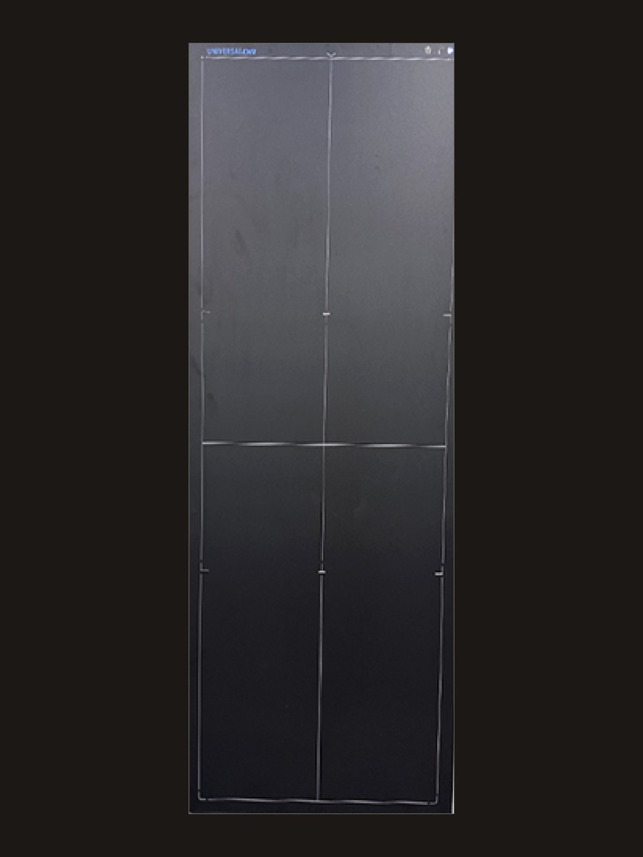
This is the panel I start with when a chiropractic room wants the full spine in one exposure. Stitching 17×17 images is still a great option for most chiropractors, and it remains in the software when a study calls for it. The 17×48 is the single-exposure path: the upright spine, a preview in under six seconds, cesium, 139 µm, 3.6 LP/mm, and 16-bit. Scoliosis and a full-spine lateral are the studies this detector is built for.
The software is the other half of the decision. Symmetry-Sync, which a lot of doctors still call Opal-RAD, already knows the work: Gonstead, Blair, Orthospinology, George's lines, Cobb, Penning, spine labeling, and stitching when a study calls for it. REALISM™ is the processing that holds trabecular detail while the exposure stays modest. For a chiropractic room that wants those tools with the long panel, this is the combination I start with.
It is a tethered detector, which suits a wall stand that stays in one place. A cable is a reliable way to run a panel that never travels between rooms. Wireless is a great fit for a detector you carry, and that is a different product. This detector is 17 × 48 inches: one exposure of the full spine. Automatic exposure detection works with most generators already on the floor, so a retrofit is a realistic path.
The panel is half of the install. The holder that goes with it is the Universal DR wall-mounted Trans-Former™. It is a full-spine grid cabinet made for this detector: 25 inches of vertical travel, a rotating pivot clutch that turns the panel 90 degrees, and a removable 8:1, 103-line parallel grid. Parallel matters on a long spine because you are not locked to one source-to-image distance (SID), the distance from the tube’s focal spot to the detector, the way a focused grid is. A focused grid is sharpest at one set distance. Parallel stays useful as that distance changes. Covering the full 48 inches takes distance too. A tube set up for a 17×17 at 40 inches will not light this panel from there. Fujifilm’s cart notes ask for about 80 to 95 inches of (SID) to capture a 50-inch field. Eighty-four inches sits inside that range, and it is the distance a chiropractic full-spine film has traditionally been made at. The tube’s anode angle decides the exact number, so it is measured in the room. The panel carries a 60-month manufacturer warranty.
20/20 Imaging is in Crystal Lake, Illinois, a division of Konica Minolta Healthcare Americas. US support is part of what I look at on the day a detector is down, along with the warranty and how the part actually ships. See the 17×48 full-spine panel, the 20/20 Imaging page, and Symmetry-Sync. If the room is ready to be measured, the 17×48 room survey is the next step.
Advantages and disadvantages
Advantages
- One exposure of the upright spine, with a preview in under six seconds.
- Cesium scintillator, 139 µm, 3.6 LP/mm, 16-bit.
- Symmetry-Sync already includes Gonstead, Blair, Orthospinology, Cobb, Penning, labeling, and stitching.
- CsI scintillator and 3.6 LP/mm resolution support lower exposure without losing diagnostic detail. Single-shot geometry removes overlap dose from stitching. No automatic exposure control — the operator's technique chart is the ALARA control.
- The Trans-Former holder travels 25 inches and turns 90 degrees, with a removable parallel grid.
- A 60-month manufacturer warranty.
Disadvantages
- Lighting all 48 inches takes roughly 80 to 95 inches of (SID), the range Fujifilm publishes for a 50-inch field. Eighty-four inches is a working distance inside that range. A 40-inch room will not cover it.

Fujifilm FDR D-EVO GL — an original long-length line, and still a great one
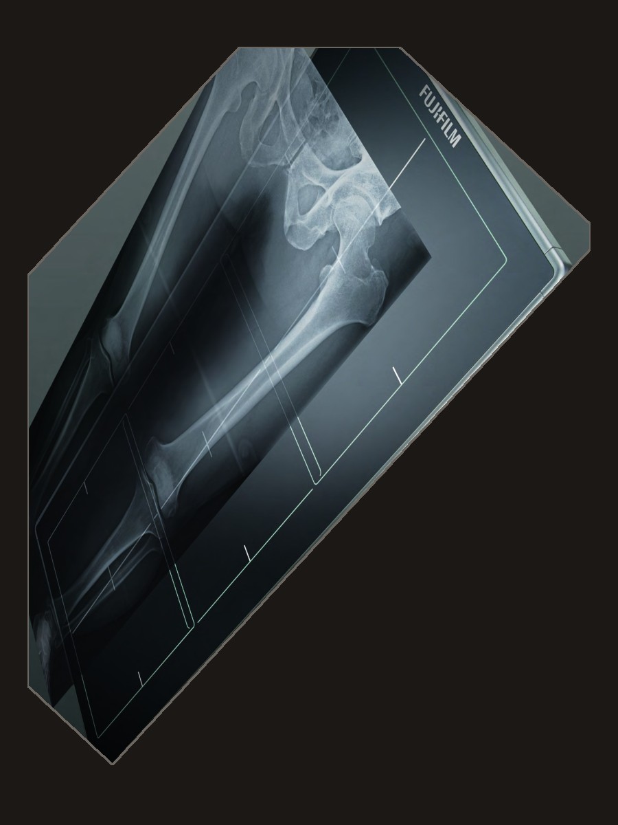
The FDR D-EVO GL is a no-brainer when the practice already works in Fujifilm, and it is a strong choice when dose tools are the priority. Fujifilm is one of the original digital radiography lines, and it firmly deserves its place on this list. The detector is 17 × 49 inches, 150 µm, 3.3 LP/mm, with a GOS scintillator and ISS (irradiated side sampling). It has its own length, its own processing, and a workflow a Fuji room already understands. If Fujifilm is new to the practice, those same tools are still the reason to look at it.
A full-length preview is 9–20 seconds, and about 6 seconds on a 17×17 segment of the same detector. Preview is how long you wait to see the image. The full cycle is about 26 seconds, or 10 on that smaller segment. Cycle time is how long until the next view can be taken. Weight is about 43 lb. The detector is about 52.7 × 22.3 × 2.0 inches. It connects with a cable to its power unit and Ethernet to the workstation, and it uses its own 120 VAC supply.
The Fujifilm toolset is a real reason to choose it. ISS is how a GOS plate makes use of the exposure: the light is read from the side the X-ray enters. Virtual Grid is Fujifilm’s published claim of up to 50% dose reduction versus a physical grid. Dynamic Visualization and the noise-reduction circuitry, listed at 1.7× DQE (a measure of how efficiently the detector uses the exposure), belong to the same story. SmartSwitch is automatic exposure detection, so most generators do not need a new trigger cable. Generator compatibility is confirmed with you before anything is ordered. Warranty is confirmed on the quote.
Fujifilm Healthcare Americas is in Lexington, Massachusetts. The parent company is in Japan. A US office matters on the day a panel is down. Fujifilm offers a shorter version of a long-view detector. Check out the G80i and Fujifilm’s many other products we sell and service.
Advantages and disadvantages
Advantages
- One of the original long-length flat panels, and still a great one: 17 × 49 inches.
- ISS, Virtual Grid, and Dynamic Visualization are a real dose and image toolkit.
- Virtual Grid claims up to 50 percent dose reduction versus a physical grid, and the 1.7× DQE noise reduction is part of the same dose story. The GOS scintillator needs more exposure than CsI to reach equivalent image quality, which is the trade for ISS.
- A no-brainer when the office already works in Fujifilm, and a strong look when it does not.
- SmartSwitch sees the exposure on most generators without a new trigger cable.
- A shorter long-view detector, the G80i, is there when 49 inches is more than the room needs.
Disadvantages
- The full cycle is about 26 seconds. Some days that pace is exactly right. Some days you will want the image sooner.
- It uses its own power supply, so the outlet at the stand is part of the plan.
- The scintillator is GOS, a powder, rather than cesium iodide. Cesium grows in needles, so the light from the exposure stays in a narrow path and more of the dose becomes image. In a powder the light scatters sideways before it reaches the pixel, which costs sharpness and usually costs dose. ISS reads Fujifilm’s plate from the side the X-ray enters, and that recovers a real part of what the powder loses. It does not make GOS into cesium.
- Fujifilm’s cart notes ask for about 80 to 95 inches of SID to capture the full 50-inch field. The room has to have that distance.

Vieworks VIVIX-S 17×51 — the longest field on this page
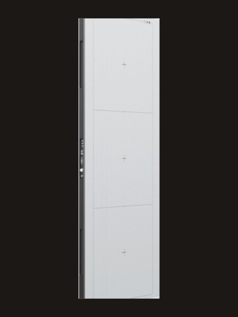
Active area is how much of the patient fits on the image. The Vieworks VIVIX-S 1751S is the longest on this page: 17 × 51 inches (430 × 1290 mm). On a tall patient, those extra inches are what keep the skull or the pelvis from being cut off. The same detector can also take a 17×34 or a 17×17 image without being moved out of the stand. Pixel pitch, the spacing between pixels, is 140 µm. Spatial resolution is 3.5 LP/mm. The full image is 3072 × 9216 pixels, at 16-bit. Sampling is close to the 2020, and the field is longer than the 17×48, the Fujifilm 17×49, the Medicatech panel, and the Delworks 17×42.
Acquisition is about 8 seconds, with a 15-second recommended wait before the next exposure. The scintillator, the material that turns the X-ray into light, is offered as cesium iodide (CsI) or Gadox. Cesium uses the exposure more efficiently. The quote names which one you are looking at, because that choice changes dose. The panel is about 19.7 kg (43 lb) and 135.7 × 53.2 × 3.0 cm. Power is DC 24 V at 3.0 A. The stated range is 40–150 kVp, which covers the energies used for diagnostic spine work. The image travels over Gigabit Ethernet. The exposure can be started with a DR trigger cable or with automatic exposure detection. Generator compatibility is confirmed for the room you have.
The hardware feature that stands on its own is multi-scan: a long spine, a shorter section, or a 17×17 region without pulling the panel out of the stand. Chiropractic measurement software is a separate choice with this detector. For a room that wants the longest field of view, this is the detector to consider.
Vieworks is headquartered in Anyang, South Korea, with US support in Vernon Hills, Illinois. That US office is who you reach for parts and a person on this continent. Warranty and the scintillator choice are both named on the quote. Service context is on the Vieworks page.
Advantages and disadvantages
Advantages
- The longest imaging area here: 17 × 51 inches, so more of a tall patient stays on one image.
- The same panel can take a 17×34 or a 17×17 without being pulled out of the stand.
- CsI option uses exposure efficiently; the Gadox alternative changes the dose math. Multi-scan modes mean 17×51, 17×34, and 17×17 from one detector, so you are not forced into a full-field exposure when a smaller view suffices.
- 140 µm and 3.5 LP/mm, in the same sampling neighborhood as the 2020.
- About 8 seconds to acquire, with a 15-second recommended wait before the next exposure.
- Cesium or Gadox, and a trigger that can be automatic or a DR cable. US support is in Vernon Hills, Illinois.
Disadvantages
- Chiropractic measurement software is a separate choice. It is worth talking through before the panel is the only decision.
- The quote should name CsI or Gadox. That choice changes how the panel uses the exposure.
- A 51-inch field needs the tube at least as far back as a 48-inch field, and a little extra room is welcome. The same 80 to 95 inch neighborhood applies.

iCRco iDR-L — the scanning long-length system
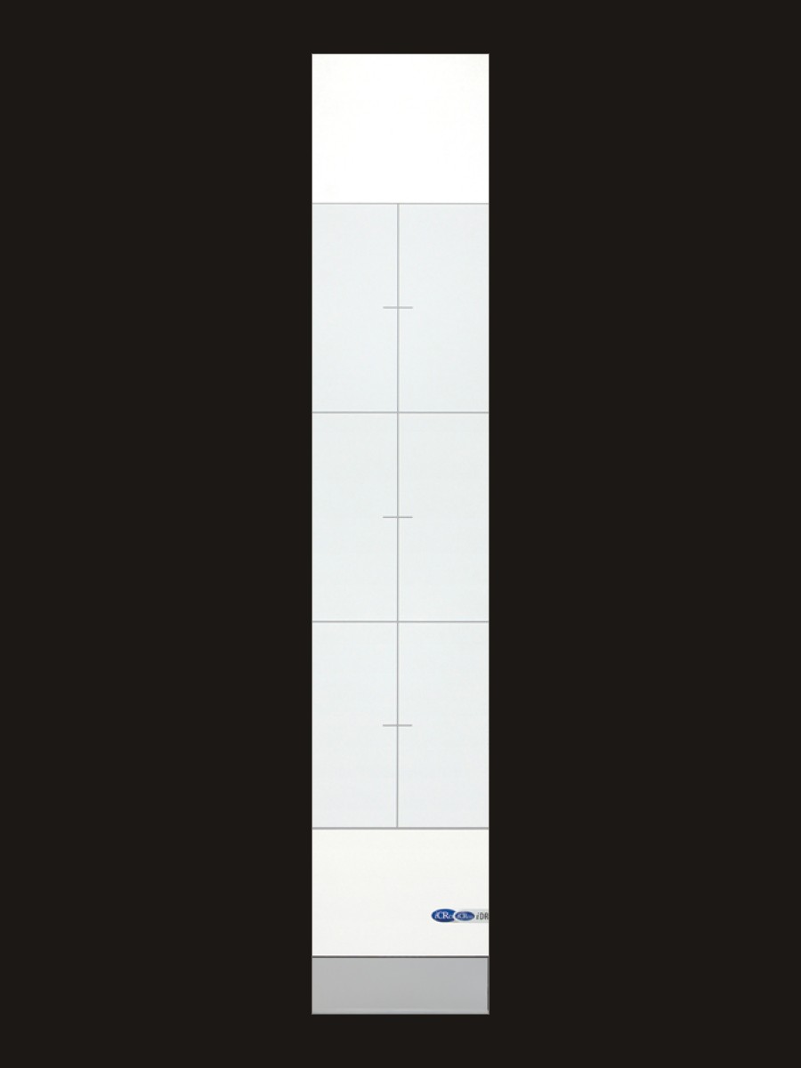
The iDR-L is iCRco’s long-length system, and it belongs in this comparison. It is a scanning phosphor-plate design, often called CR, built as a tall wall stand rather than a flat-panel detector. A flat panel makes the image during the exposure. A scanning system reads the plate afterward. That is an earlier technology, and the published numbers are still excellent. The scan area is 17 × 51 inches, in the same range as the longest flat panel on this page. If you are deciding between a scanner and a flat panel, both are here so you can compare them with the facts in front of you.
Selectable scan modes are 100, 155, and 200 µm, published as 5.0, 3.1, and 2.8 LP/mm. Five line pairs is the highest published resolution in this comparison. That finest mode takes about 55 seconds while the scanner moves across the receptor. The choice is a high-detail read that takes longer, versus a flat-panel preview that appears in seconds. Both are legitimate ways to make the image. Laser spot size and plate characteristics shape a CR image the same way pixel pitch shapes a DR image.
The wall stand is about 100 lb. It is the imaging system itself, not a panel that drops into a stand you already own. A trigger cable to the generator tells the scanner that an exposure happened. Generator compatibility is confirmed before the system is ordered. Chiropractic measurement software is chosen with you, so the tools on the screen match how you read a spine.
iCRco builds in Goleta, California, with support listed Monday through Friday, 8am–6pm Pacific. A US manufacturer is a practical advantage for hours, shipping, and parts. For a typical chiropractic full-spine room that wants a flat-panel preview and measurement tools in the same system, I start with the 2020. If this scanning wall stand is what you want, tell me why. That is a good conversation, and the iDR-L is quoted with the same care as the flat panels.
Advantages and disadvantages
Advantages
- Published resolution up to 5.0 LP/mm in the finest scan mode. Those numbers are excellent.
- A 17 × 51 inch scan area, in the same long range as the longest flat panel here.
- No published DQE or dose-reduction claims. The 100-micron scan mode at 5 LP/mm is the finest resolution here, but finer sampling does not by itself lower patient dose — it raises the bar for what the generator must deliver.
- Built in Goleta, California, with weekday support on Pacific time.
- It is the wall stand, so you are looking at a complete scanning system rather than a panel plus a cabinet.
Disadvantages
- A flat panel makes the image during the exposure. Nothing inside it has to travel. The iDR-L stores the exposure on a phosphor plate, then a scanner inside the stand moves along that plate and reads it with a laser. That carriage is why a full-resolution image takes about 55 seconds.
- Those moving parts are in the image path: the laser, the drive that carries it, and the plate itself. Plates scratch and pick up dust. When the scan alignment drifts, or one of those parts wears, the image shows it, and the stand needs a service a solid-state panel never asks for.
- The 5.0 LP/mm figure is the slowest, finest mode. The faster modes are published at 3.1 and 2.8 LP/mm. You are choosing between a slower high-detail scan and a quicker read, not getting both at once the way a flat panel does.
- It uses a trigger cable to the generator, and the stand is about 100 pounds.
- The exposure still has to cover a long plate, so the tube distance question is the same as it is for the flat panels.

Medicatech Venu 1748 — full spine and long bone, wired or Wi-Fi
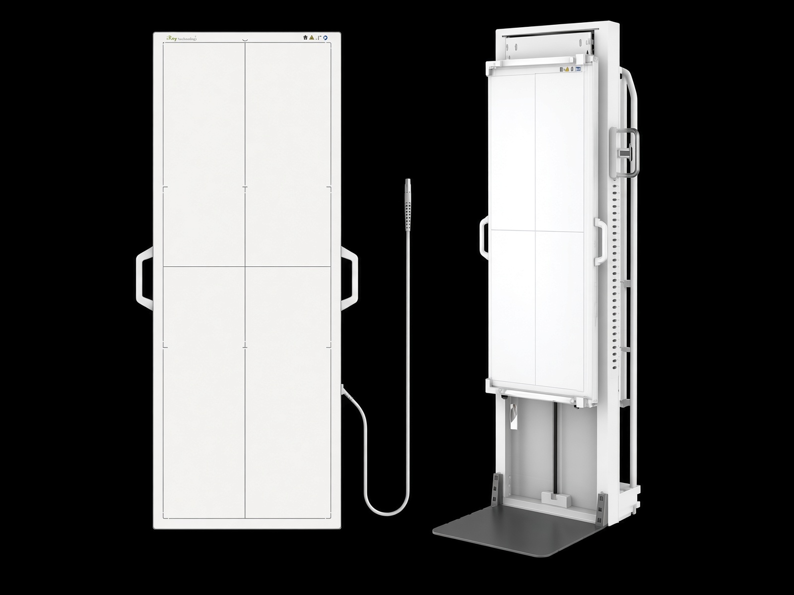
This column is the detector in Medicatech’s full-spine and long-bone brochure, the Venu 1748. It is not a different Medicatech long-length panel. If a sheet in front of you says MX, or names a 17×51 or 17×34 IGZO detector, that is another product and it is not this column. The photo matches the brochure: a tall white panel with side handles, marked iRay, and a floor stand beside it.
Both models are amorphous silicon with a cesium iodide scintillator. Active area is 425 × 1208 mm. That is 16.7 × 47.6 inches. The brochure prints millimeters, not inches, so the inch figure is the conversion. Pixel size is 139 µm. Medicatech publishes spatial resolution at 3.4 lp/mm. The Nyquist math from 139 µm is 3.6, and I left their published 3.4 in the matrix. Bit depth and the pixel matrix are not on this brochure. Outer size is 1271 × 487 × 21 mm. Pre-image time is 3 seconds. Full image time is 8 seconds. Power is 27 watts. The wired 1748V weighs 17 kg, about 37.5 pounds. The 1748V-WF weighs 18 kg with the battery, about 39.7 pounds, and the battery is listed at 3 hours on a 90-second cycle.
The wired model sends the image over GigE. The WF model is GigE or Wi-Fi. Trigger is software, with AED optional, so the quote has to say whether your generator needs that option. The brochure’s claim for the clinical job is one exposure of the full spine or the whole lower extremity, instead of stitching several shots. Voyance is the acquisition software: automatic optimization, more than one language, and optional iGrid, which they describe as removing light scatter. iGrid is software. A physical grid ratio is not on this brochure. Voyance is not a Gonstead or Blair package. If the room reads that way, the software choice is a separate conversation, the same way it is with Vieworks.
Two holders are on the sheet. The rolling cart is the DR-1748A: 1102 × 680 × 151 mm, a cross arm that adjusts from 350 to 1350 mm, armrests that move 100 mm up or down, and a 90-degree rotation. The floor stand is the DR-1748D: 595 × 500 × 1906 mm, detector vertical travel 1300 to 1770 mm, horizontal travel 575 to 1375 mm, the tray rotates 90 degrees, and the casters unfold 30 degrees. That is a yes on the portable-stand row. Warranty, kVp range, and a headquarters city are not on the brochure. Medicatech USA lists support since 2005, phones +1 800-817-5030 and +1 949-679-2881, and medicatechusa.com. I am not guessing a city.
Advantages and disadvantages
Advantages
- One exposure for a full spine or a whole lower extremity, on a cesium panel at 139 µm.
- A wired model and a Wi-Fi model. Preview in 3 seconds, full image in 8.
- CsI scintillator at 139 microns, consistent with the efficient end of the dose spectrum. iGrid removes light scatter that would otherwise require a grid and its attendant dose. Portable cart and floor stand options mean the panel can leave the room when ALARA calls for a different geometry.
- A rolling cart and a floor stand are both on the brochure, so the panel does not have to live in one wall cabinet.
- About 37.5 pounds wired, which is lighter than the 43-pound wall panels on this page.
Disadvantages
- Active area is about 47.6 inches, shorter than the Vieworks 51 and a little shorter than the 2020 48. A tall spine can still get cropped.
- Bit depth, pixel matrix, warranty, kVp range, and a physical grid ratio are not on this brochure.
- AED is optional. Confirm the trigger against the generator before anyone orders the panel.
- The published spatial resolution is 3.4 lp/mm, under the 3.6 the pixel pitch would suggest. I am not rounding their number up.
- Voyance is acquisition software. It is not the chiropractic measurement set I start a chiropractic room with.

Delworks LLI — the lighter panel that can leave the wall
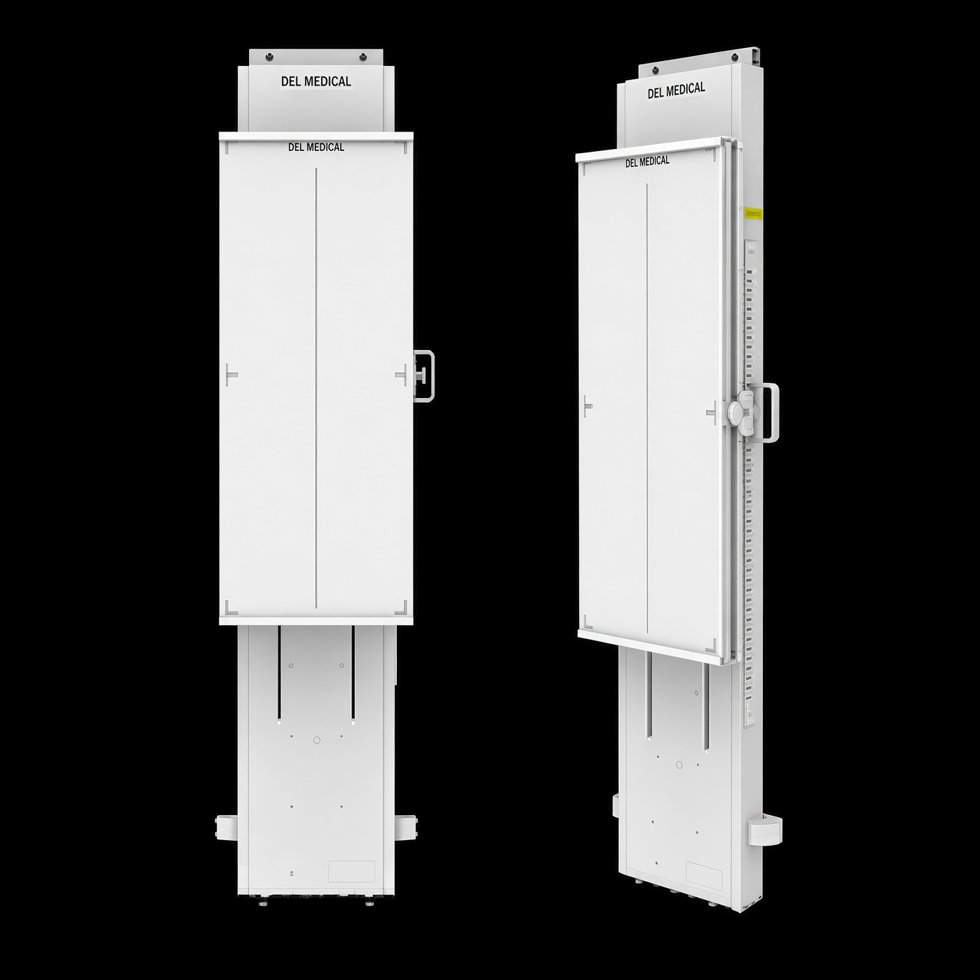
The DELWORKS LLI is UMG / Del Medical’s long-length flat panel. The current brochure is #9000-DELWORKSLLI, Rev. C. The specification table is what I used for the matrix. Active area is 17 × 42 inches (43 × 107 cm). That is the shortest flat panel on this page. It will cover many spines and a long leg study, and it will not cover a tall standing spine the way a 48-, 49-, or 51-inch panel does. The reason to look at it anyway is everything else on the sheet: one exposure, no stitch, 22 pounds, and holders that are actually mobile.
The specification table says cesium and amorphous silicon, 139 µm, 16-bit, 3.6 LP/mm, matrix 7680 × 3072 (24 megapixels), acquisition 3 seconds, 40–150 kV, GigE and Wi-Fi, 20 watts, and 22 pounds (10 kg) with the battery. Housing is carbon and aluminum. Water tightness is IPX3. Outer size on that table is 111.5 × 47.1 × 1.5 cm. The drawing on the same page marks about 44 × 18 × 0.8 inches. I am not blending those into one thickness. The highlights box says a 9-second image display and 140-micron pixels. The 2021 white paper says ready for viewing in 9 seconds and 24 pounds. Where those disagree with the specification table, the table wins for the matrix, and the other figures stay labeled as what they are.
One features paragraph says the panel “contains a Gadox scintillator,” and the low-dose paragraph on that same page says Cesium Iodide. The specification table says Cesium. I am using Cesium. If a quote you already have says Gadox, ask them to match it to the current table before you sign.
AED is how it sees the exposure, and Del says it works with any generator. I still confirm the generator. Wireless is 802.11ac, talking to DELWORKS 3.0. Two lithium-ion batteries take 2 to 3 hours to charge, then 650 exposures or 11 hours on standby. The wired option is a 15-foot magnetic cable, and that cable charges the panel too. You can collimate to a smaller view and the software auto-shutters, which is how the same detector does a routine study. Optional filters — lateral cervical, AP/PA scoliosis, and leg length — mount magnetically in the collimator rails. DELWORKS 3.0 has a positioning guide, a deviation index, and measurement tools. It is not a named Gonstead or Blair package.
Portable stand is a yes, and it is the clearest yes on this page. The Mobile Positioning Holder does upright, supine under-table, and cross-table work, including the operating room: telescoping arm, piston travel, locking casters. The Mobile Vertical Holder lowers the image to 1.75 inches from the floor and turns the panel from portrait to landscape. The VS50 is the wall stand when you want it to stay put: under 7 inches from the wall to the receptor, four-point wall mount, no floor drilling, no power, lead backer, 37.5 inches of vertical travel. The optional grid is 17×42, 8:1, 103 lines, 44 inches to infinity, so the same grid is usable at a routine distance and at a long-spine distance. A supine cover is rated at 500 pounds. Del’s offices on the brochure are Harrison, New York, and Bloomingdale, Illinois.
Their 2019 launch language, in the white paper, calls this the world’s first monolithic long-length DR detector. That is their sentence. The OTC18S from 2021 is a stitching room that uses a standard-size detector. It is in the history section so you know the name. It is not this panel.
Advantages and disadvantages
Advantages
- 22 pounds on the current spec table, with Wi-Fi or a 15-foot cable that also charges.
- Two mobile holders, plus a wall stand that needs no power and no floor drilling.
- Del's own white paper recommends attenuation filters under ALARA for scoliosis and leg-length views, and the 22-pound weight makes alternative positioning practical. One exposure, no overlap dose — but the 17×42 field is the shortest here, so taller patients may still need a second view.
- Cesium, 139 µm, 3.6 LP/mm, 16-bit, one exposure. Collimate down for a routine view on the same panel.
- Optional parallel grid, 44 inches to infinity, and optional scoliosis and leg-length filters.
- AED, and a US company with offices in New York and Illinois.
Disadvantages
- 17 × 42 inches is the shortest flat-panel field here. A tall full spine can be cropped.
- The brochure does not agree with itself in two places. Spec table: Cesium, 139 µm, 3-second acquisition, 22 lb, 1.5 cm thick. Elsewhere: a Gadox sentence, a 140-micron highlight, a 9-second display, and a drawing at 0.8 inch thick. The 2021 white paper says 24 lb. The matrix follows the spec table.
- DELWORKS is not the chiropractic analysis set. If you read Gonstead or Blair, say so before the software is treated as settled.
- “World’s first” and “no other long-length detector is as portable” are Del’s marketing lines. The weights on this page are here so you can compare them yourself.
How To Get Started Now
You know your patients and how the day actually runs. I can walk you through what is on this page, and you can tell me what fits your room and what does not. Tell me what you already like, what is getting in the way, and what a good full-spine image has to do for you. The more of that I hear, the better I can help.
A few practical details make the quote accurate. Generator compatibility is one of them. Some of these systems see the exposure on their own. The iCRco uses a trigger cable. Fujifilm uses its own power supply. Medicatech’s AED is an option, and the Wi-Fi model is the one with a battery. Delworks is wireless or a charging cable, and it is the panel with two mobile holders. None of that is a mark against the equipment. It is just how each one connects to the room you already have. Wall space, ceiling height, and the stand are measured with you, not guessed. If the panel has to roll, say that first. Only Medicatech and Delworks publish a portable holder on the sheets this page uses.
Prices stay off this page. A long-length room is quoted for your generator, your stand, and the software you want to read with. Call 720-600-3117 or send a note. The chiropractic page covers the rest of the upright room — straight-arm, DDR, and a smaller detector when a 17×48 is more panel than the practice needs.
Frequently Asked Questions (FAQ)
What is a long-length or full-spine DR panel?
It is a single detector tall enough to cover a standing spine in one exposure. Clinics also call it a long-view or long-form panel. Many offices still make a full spine by stitching 17×17 images, and that remains a great way to work. A long-length panel is the one-exposure version of the same study.
Is stitching three 17×17 images the same as a long-length detector?
Stitching is still a great option for most chiropractors. It joins separate 17×17 exposures in software, and a lot of offices do excellent work that way. A long-length detector is a different choice: one exposure and one image, when the room is ready for a single tall panel. One is not a failure of the other.
Which long-length panel fits most chiropractic offices?
For most chiropractic full-spine rooms, I start with the 2020 Imaging Universal 17×48. It is a cesium flat panel, preview is under six seconds, and Symmetry-Sync is built for chiropractic measurement. Generator compatibility is confirmed as part of the quote, and what you already like about your room is part of that conversation.
When would Fujifilm or Vieworks be the better fit?
Fujifilm FDR D-EVO GL is a natural fit when the room already uses Fujifilm, and when dose-reduction tools are the priority. It is one of the original long-length lines, and still a great one. Vieworks VIVIX-S 17×51 has the longest imaging area here, 17 by 51 inches, so more of a tall patient fits on one image. The same panel can also take a 17×34 or 17×17 view without being moved.
Which of these have a portable stand?
Medicatech and Delworks, on the documents this page uses. Medicatech offers a rolling cart (DR-1748A) and a floor stand (DR-1748D) for the Venu 1748. Delworks offers a Mobile Positioning Holder and a Mobile Vertical Holder, plus the VS50 wall stand if the panel stays on the wall. The 2020 holder on this sheet is the wall-mounted Trans-Former. The iDR-L is the wall stand. Fuji and Vieworks do not publish a portable long-length stand on the sheets I used, so I am not calling that a no.
Is the Delworks LLI long enough for a full spine?
It is 17 by 42 inches. That is shorter than the 2020 at 48, Fuji at 49, and Vieworks and the iCRco scan area at 51. Forty-two inches covers a lot of spines and it covers long legs, and it will crop a tall standing spine that the longer panels still hold. The reason it is in this comparison is the other half of the spec: 22 pounds, wireless or a short charging cable, and holders for upright, table, and cross-table work. Del’s 2021 white paper also recommends about 100 inches of source-to-image distance when you want the whole panel covered.
Is the iCRco iDR-L a flat-panel DR detector?
No. It is iCRco’s scanning phosphor-plate wall stand, with a 17×51 scan area. The published 5 LP/mm figure is its finest scan mode, and those numbers look excellent. A full-resolution read is about 55 seconds because the scanner travels the plate. It is an earlier technology than a flat panel, and it is a fair system to compare when you want both ways of making a long-length image in front of you.